Case Studies
Case1
A 30-year-old woman, weighing 48kg. She presented with heaviness in the lower abdomen since 6 months and also had oligomenorrhea and dysmenorrhea. She is an otherwise healthy person.
Transabdominal USG performed showed a 7.9 x 7.4 cm sized anterior myometrial fibroid. MRI showed a hypointense fibroid on T2 weighted images measuring 11.5(TR) x 11.5(SI) x 9.1(AP) cm (Fig. 1).
Ablation of the fibroid was carried out using High Intensity Focussed Ultrasound under MRI guidance. The entire procedure lasted about 2½ hours, after which intravenous gadolinium based contrast was administered, and a non-perfused volume of 85-90% was obtained (Fig. 2). The patient tolerated the procedure well.
She had abdominal discomfort for a day, but she could resume her daily activities normally
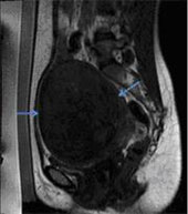
Fig. 1 – T2 weighted sagittal image showing a large fundal hypointense fibroid (blue arrows), with a central area of hyperintensity, representing internal degeneration (red arrows).
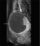
Fig. 2 - Post Contrast Fat Saturated T1 weighted sagittal image showing a large internal area of non-perfusion (arrows), which is the ablated area
Case2
A 39-year-old married woman, with two children, weighing 65kg. She had a transverse lower abdominal scar due to a prior Caesarian section delivery. She presented with 2-3 episodes of severe abdominal pain, superimposed on continuous dull lower abdominal pain since 2 months.
Abdominal CT scan showed a fundal fibroid measuring 12.9 x 11.0 cm, with areas of internal degeneration. MRI showed a hypointense fibroid on T2 weighted images, measuring 12.9(TR) x 14.0(SI) x 9.3(AP) cm, with a large central area of degeneration within (Fig. 1).
She had an intrauterine contraceptive device (IUCD), which was removed before the procedure. Ablation of the fibroid was carried out using High Intensity Focused Ultrasound under MRI guidance. The entire procedure lasted about 2 hours, after which intravenous gadolinium based contrast was administered a non-perfused volume of 75-80% was obtained (Fig. 2). The patient tolerated the procedure well.
She had abdominal pain for a day, which was relieved with analgesics. She also had nausea for 2-3 days after the procedure but she could resume her normal daily activities
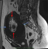
Fig. 1 – T2 weighted sagittal image showing a large fundal hypointense fibroid (blue arrows), with a central area of hyperintensity, representing internal degeneration (red arrows).
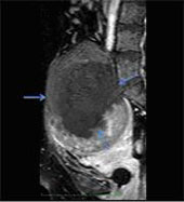
Fig. 2 - Post Contrast Fat Saturated T1 weighted sagittal image showing a large internal area of non-perfusion (arrows), which is the ablated area.
|