| Coronary
Artery Imaging
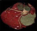
Coronary and Cardiac Anatomy
Basic anatomy
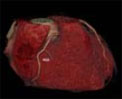
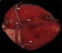
Heart
It consists of four chambers and
the vessels that enter and leave.
Blood from the body enters the right atrium (RA) through
the SVC and IVC. It then goes through the tricuspid
valve (TV) into the right ventricle (RV). The RV pumps
blood through the pulmonary artery (PA) into the lungs.
From the lungs, the blood goes via the pulmonary veins
(PV) into the left atrium (LA) and via the mitral
valve (MV) into the left ventricle (LV). The LV pumps
blood into the body through the aorta (AO), passing
through the aortic valve (AV).
All the anatomy shown here are from
images obtained at our institute.
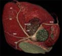
Coronary
There are four major coronary arteries.
RCA
The right coronary artery (RCA)
arises from the right coronary sinus of the aorta.
It courses in the atrio-ventricular groove between
the RA and RV and reaches the base (crux) of the heart.
It mainly supplies the RV and parts of the septum
and basal LV. In 85% of patients, it is the dominant
artery, which means that it gives off the posterior
descending artery (PDA) that supplies the inferior
wall of the LV.
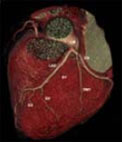
LM
The left main is a short vessel
that arises from the left coronary sinus of the aorta
and then branches into the left anterior descending
(LAD) and circumflex (CX). It may be very short or
long (upto 2cm) or sometimes absent. Disease in the
LM can be devastating.
LAD
It arises usually from the LM, but
sometimes directly from the aorta. It courses anteriorly
and inferiorly in the inter-ventricular groove between
the RV and the LV and supplies most of the anterior
wall of the LV upto the apex and the septum. It has
septal and diagonal branches, which are often large.
CX
It arises usually from the LM,
but sometimes directly from the aorta. It courses
in the left AV grove between the LA and LV and goes
towards the base (crux) of the heart. In the majority
of patients who have right dominance, the distal CX
is hypo plastic, thin or absent. In 8-10% of patients,
there is left dominance, where the PDA arises from
the CX. In about 5-10% of patients, there is co-dominance,
where the PDA arises from the RCA, but another artery,
the PL, arises from the CX. The CX supplies a large
part of the lateral and sometimes the inferior wall
of the LV and has obtuse marginal branches.
|