| Functional
Assessment
Routine functional assessment is
now being offered for the first time on a CT scanner
on the 64-slice CT. Though it has been possible in
the research setting, clinical utility has not been
forthcoming until now. Routine functional assessment
is now being offered for the first time on a CT scanner
on the 64-slice CT. Though it has been possible in
the research setting, clinical utility has not been
forthcoming until now.
In functional assessment, the following
parameters are assessed.
Wall motion
As with echocardiography and MRI,
it is possible to reconstruct the images in multiple
phases and then generate cine images to view the contractility
of all the segments of the myocardium. Though the
data and literature is still sparse (since this is
a new technique), its use can be extrapolated to the
data existing for echocardiography and MRI. We assess
for the presence of reduced contractility or absent
contractility in different segments of the LV. This
complements the coronary artery study.
For example, if the anterior wall
and septum are hypo kinetic, in a patient with a stenosis
of the LAD, seen on the coronary CT study, the systolic
dysfunction makes it almost certain that the lesion
is significant and needs to be treated. In fact if
systolic dysfunction is seen, it even obviates usually
the need for a stress-thallium or a stress-perfusion
MRI examination, since perfusion changes occur earlier
in the ischemic time-line. By the time systolic 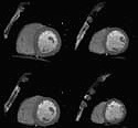 dysfunction
occurs, there will always be perfusion changes. dysfunction
occurs, there will always be perfusion changes.
Short axis cine of
a patient with good contractility and wall thickening
of the segments
Ejection fraction
By measuring the end-diastolic and
end-systolic cavity sizes, the volumes and consequently
the ejection fraction can be measured. Though again
the data is sparse, a few studies done recently have
shown that CT underestimates by 5-10% the ejection
fraction as compared to MRI, the gold standard. This
should be taken into account when interpreting the
results.
|